Pes Varus Correction in a Dachshund: Planning, Guides and Fixation
Pes varus is a deformity of the distal tibia seen in Dachshunds, causing a bow-legged appearance, and can be associated with lateral patellar luxation. If left untreated, clinically significant deformity can contribute to persistent lameness, discomfort and abnormal joint loading, with the potential for osteoarthritis over time.
For this case, Vet Forge collaborated with Dr Vaughan Moore at SASH North Ryde to plan the correction and develop patient-specific surgical guides, tailored to the limited space available for guide positioning and implant placement.
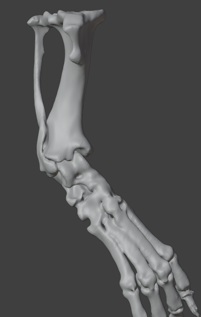
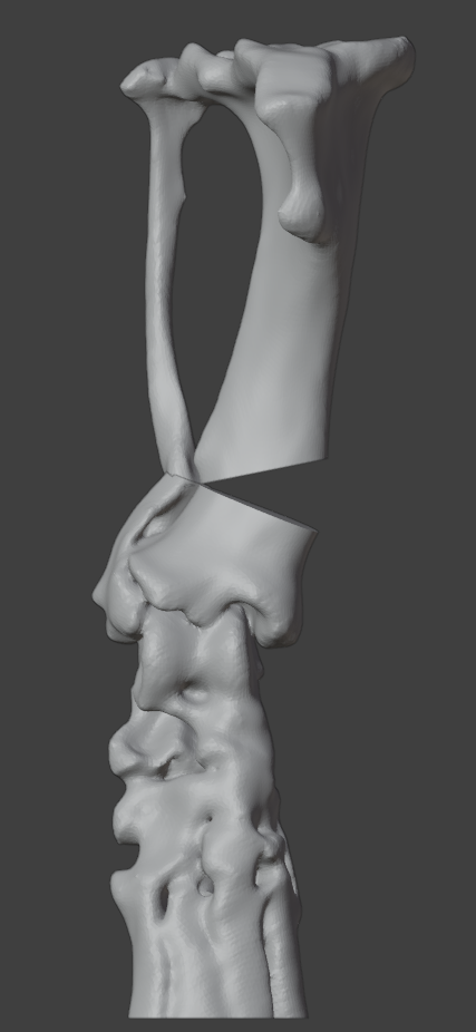
CAD Planning: Making Space for the Guide and Implant
A key planning challenge was the very distal location of the centre of rotation of angulation (CORA), close to the articular surface. This left a small distal bone segment to accommodate both the guide footprint and implant fixation.
An opening wedge osteotomy was planned, with the guide and implant positions considered together. As the main implant would occupy the medial surface of the tibia, we selected the dorsal surface for the guide footprint. This allowed the reduction guide to remain in position while the medial implant was placed and secured.
The reduction guide also incorporated a spacer to maintain the required opening wedge. This was designed to prevent the gap from collapsing during fixation, especially if the fibula fracture inadvertently.
Together, the dorsal guide position and spacer allowed the correction to be supported while preserving access for implant placement.
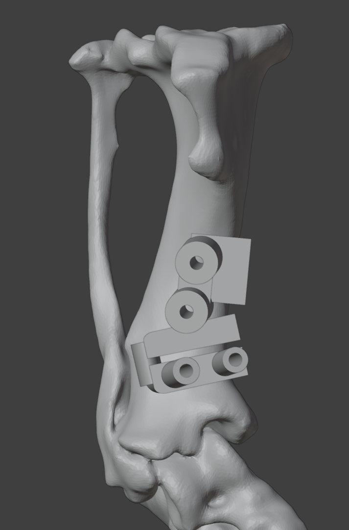
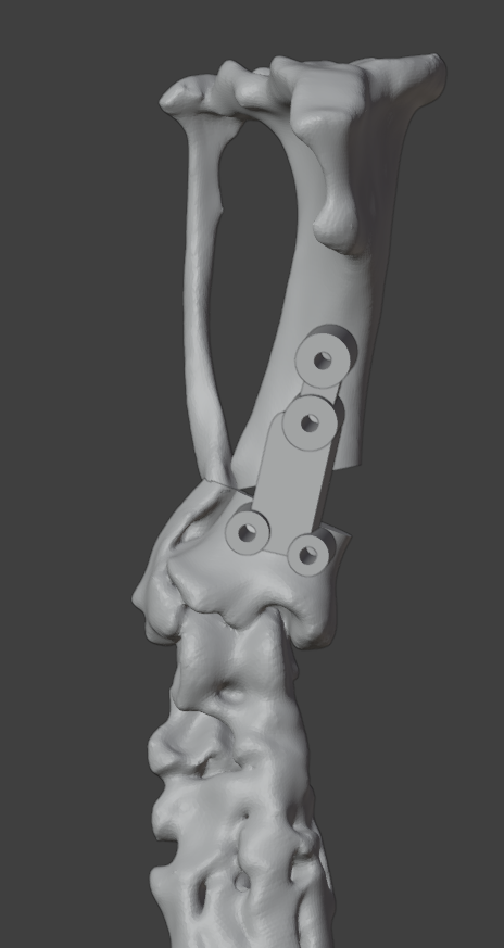
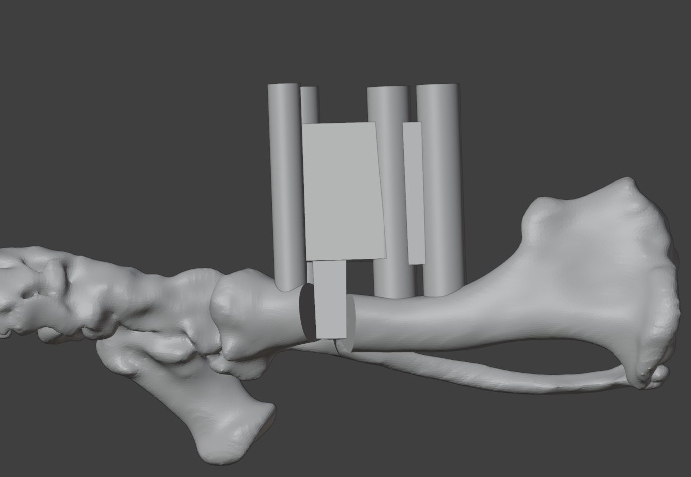
CAD images: planned osteotomy and correction, dorsal guide footprint, and reduction guide with its opening wedge spacer.
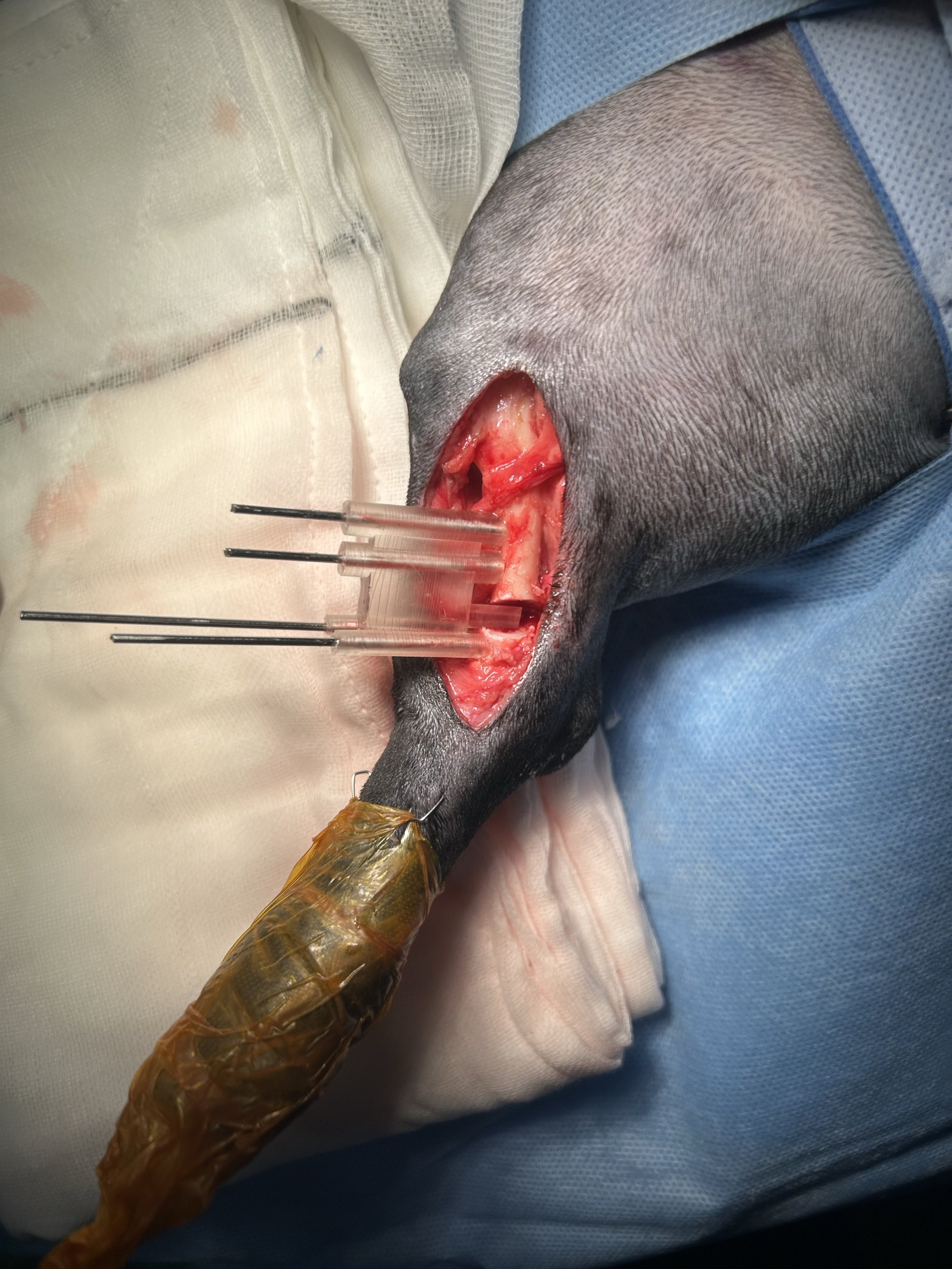
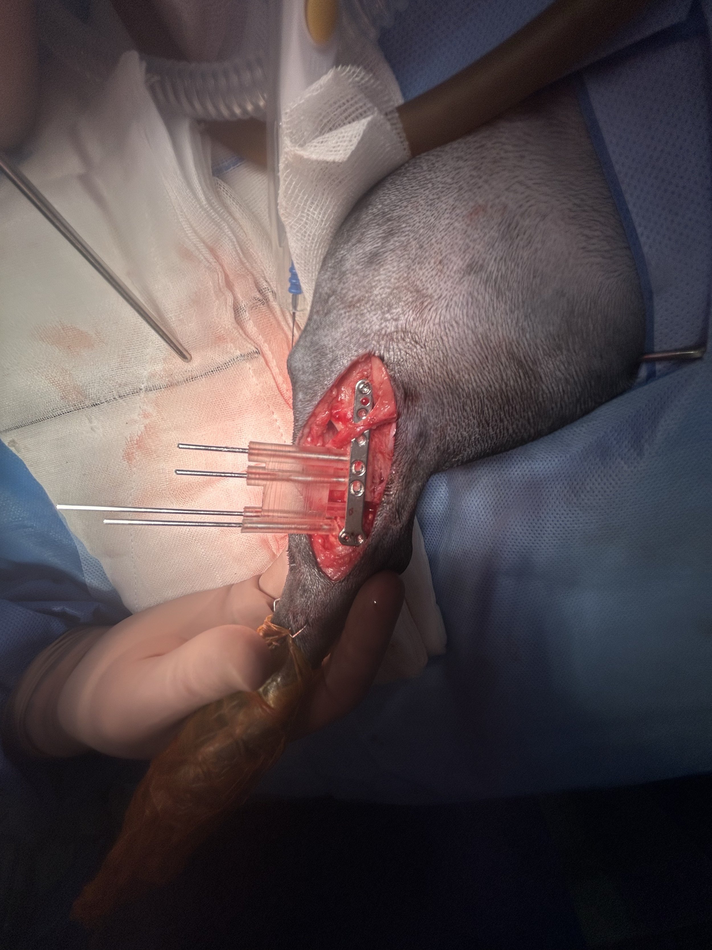
Intraoperative images: guide fit, reduction guide and spacer positioning, and medial implant placement.
Intraoperative Images: Maintaining the Opening Wedge
The intraoperative photographs document the guide fit and implant placement.
The dorsal position kept the reduction guide clear of the medial implant, allowing it to support the correction throughout fixation. The incorporated spacer maintained the planned wedge opening while the implant was positioned and secured, reducing reliance on the intact fibula to hold the gap.
This design addressed a practical challenge of opening wedge correction: maintaining the required alignment while working within the limited space around the distal tibia.
Preoperative Limb Appearance and Postoperative Radiograph
The preoperative limb photographs document the presenting deformity, while the postoperative radiograph records the osteotomy configuration and implant position following correction.
These images bring together the clinical presentation and surgical management, complementing the CAD planning and intraoperative photographs shown above.
Thank you to Dr Vaughan Moore and the SASH North Ryde team for collaborating on this case and sharing the surgical images.
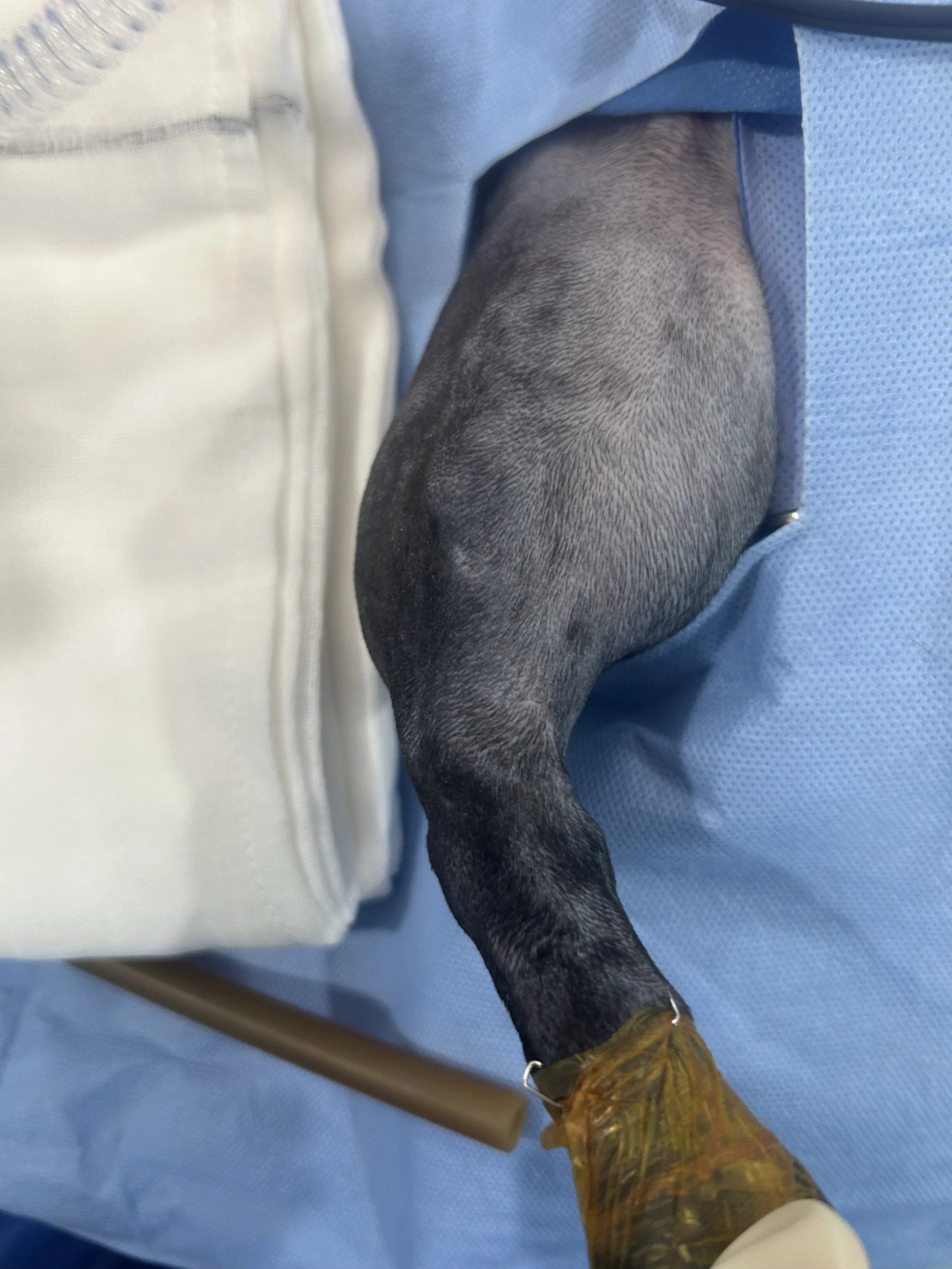
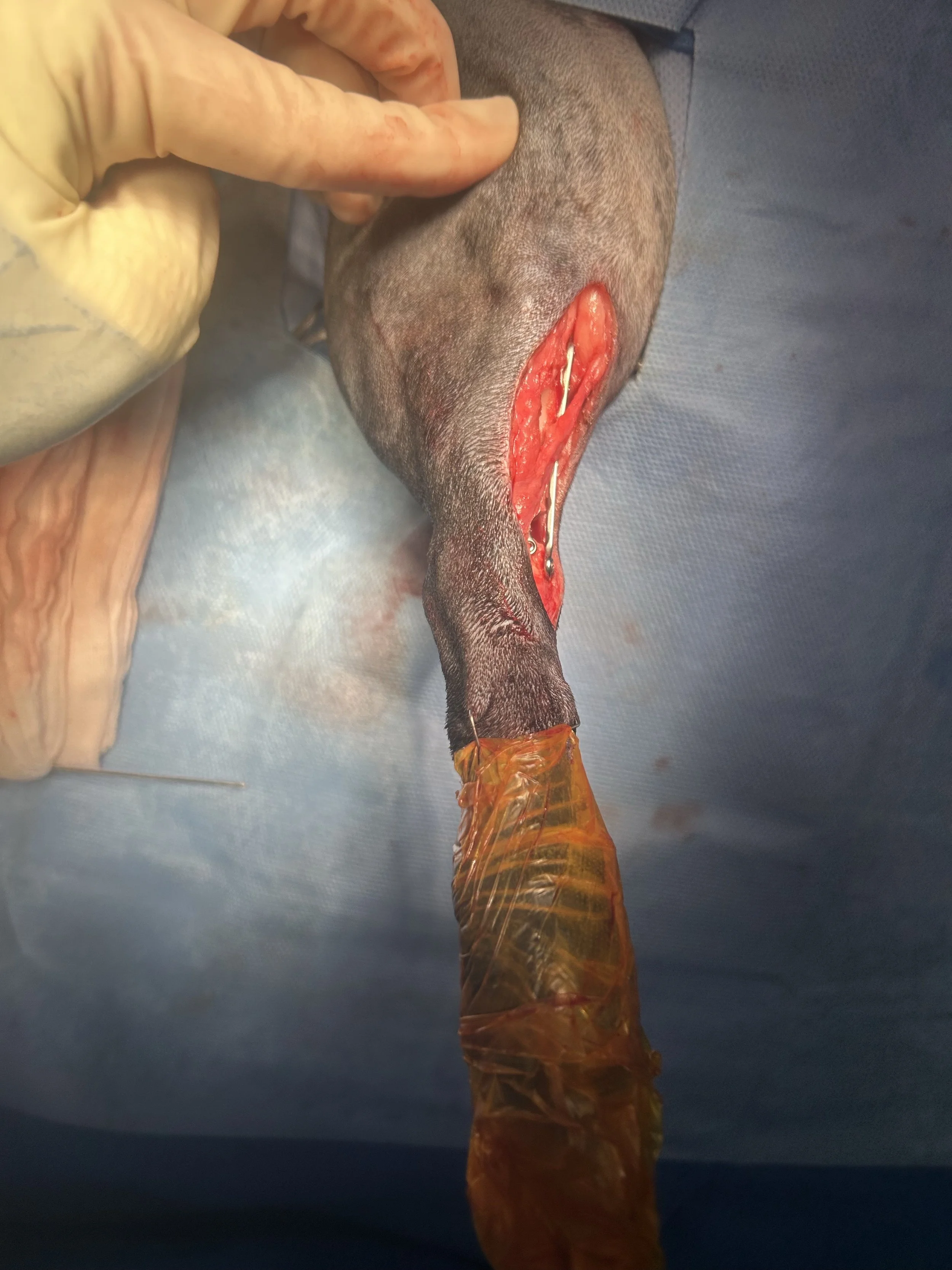
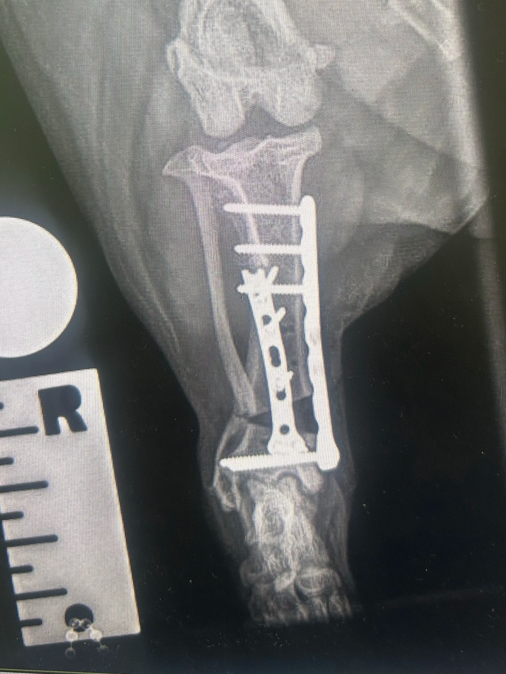
Images: preoperative limb appearance and postoperative radiograph following correction and fixation.