Patient-specific planning and guided transcondylar screw placement for humeral intracondylar fissure
Humeral intracondylar fissure (HIF) is a partial or complete fissure through the humeral condylar isthmus. If left untreated, this may result in thoracic limb lameness and an increased risk of condylar fracture.
Accurate transcondylar screw placement requires a trajectory that remains centrally positioned within the condyle while avoiding articular penetration. Freehand placement can be technically demanding, and we typically utilise intraoperative fluoroscopy to assess and refine the trajectory. Variation in condylar morphology, particularly in HIF-affected spaniels, adds complexity to identifying the optimal drilling axis, even with fluoroscopic guidance.
Archie presented to Small Animal Specialist Hospital Gold Coast with left thoracic limb lameness. Previous CT imaging demonstrated bilateral HIF and humeroulnar incongruity. CT-based planning and patient-specific instrumentation were used to facilitate bilateral transcondylar screw placement during a single anaesthetic episode.
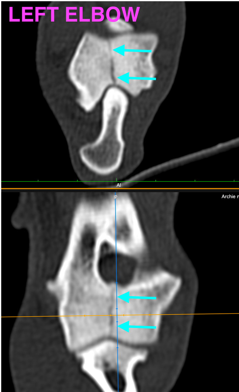
Image: CAD assessment of the proposed left transcondylar screw trajectory, demonstrating its position within the condyle and orientation relative to the fissure
CAD-planned screw trajectories
Archie’s preoperative CT data were used to generate three-dimensional bone models for planning in computer-aided design (CAD) software.
The proposed screw trajectories were assessed in multiple planes to optimise central positioning within each humeral condyle and orientation perpendicular to the fissure plane. This allowed the entry and exit points, surrounding bone margins and relationship to the articular surfaces to be evaluated before surgery.
Screw lengths were determined from the CT-derived bone models, incorporating implant sizing into the preoperative plan.
Patient-specific guide design using CAD software
Following trajectory planning, patient-specific drill guides were designed in CAD software to transfer the digital plan to the surgical site.
The bone-contacting surface was designed to conform to the local humeral anatomy, with the guide channel aligned to the planned drilling axis. This linked the guide’s anatomical fit to the intended screw entry point and trajectory.
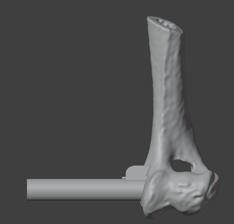
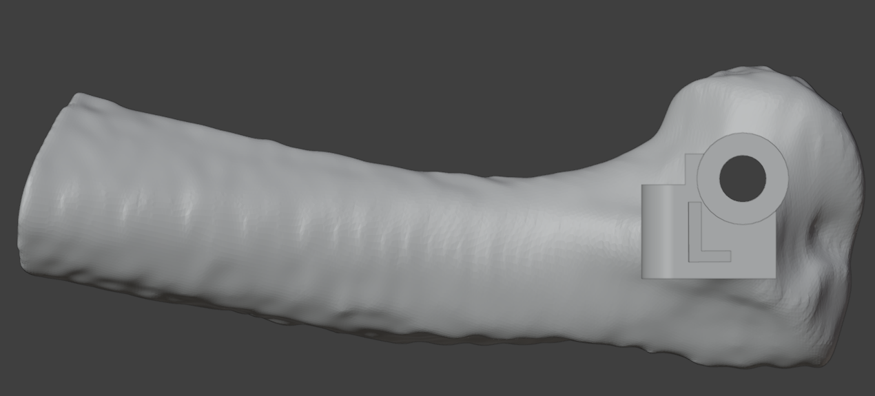
Image: CAD design of the left patient-specific drill guide, showing its relationship to the humeral surface and planned drilling trajectory.
Surgical treatment and immediate postoperative CT
Bilateral transcondylar screw placement was performed during a single anaesthetic episode using the patient-specific drill guides, without intraoperative fluoroscopy. Distal ulnar ostectomy (DUO) and interosseous ligament (IOL) transection were also performed to address the humeroulnar incongruity.
Immediate postoperative CT was obtained to assess screw positioning within each humeral condyle and the relationship to the fissure and adjacent articular surfaces.
Video: Immediate postoperative CT of the left elbow following guided transcondylar screw placement.
Eight-week follow-up
At the eight-week recheck, Archie was ambulatory without observable lameness. A subtle reduction in left elbow extension remained.
Follow-up CT confirmed progressive osseous union of the fissure
Thank you to the team at SASH Gold Coast for the opportunity to collaborate on Archie’s case and for sharing his clinical progress and follow-up imaging.
Video: Eight-week postoperative CT of the left elbow, demonstrating progressive osseous union of the humeral intracondylar fissure.